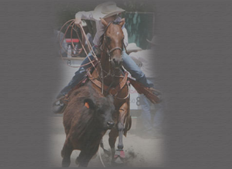
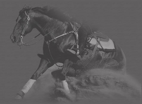
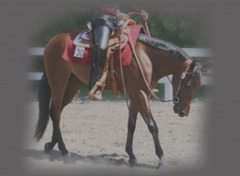
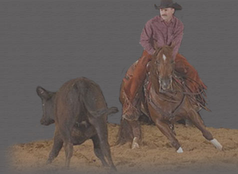
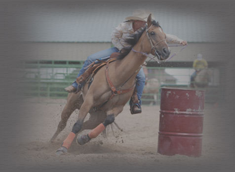
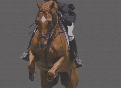
Sports Medicine
A majority of Dr. Rhoads' practice is devoted to evaluating the equine athlete for poor performance or lameness. Dr. Rhoads has extensive experience in examining horses performing a variety of disciplines. His experience as an exhibitor and horseman also provides a unique insight and ability to communicate with trainers and owners. Examinations generally consist of these components:
-
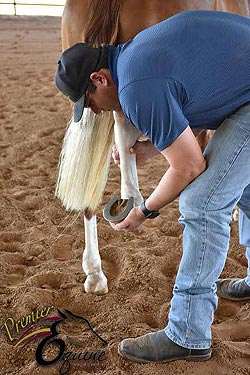
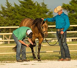
Performance/lameness
examination: The horse is examined at a walk and jog
either on a lunge line or free within a round pen. In
instances where the problem appears or feels worse under
saddle, the horse is examined under saddle. The horse is
systematically palpated over the neck, back, and all 4
limbs. Flexion tests are performed on all 4 legs on all
horses, so that subtle secondary or “compensatory”
issues may be found.
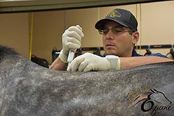
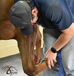
- Diagnostic anesthesia: When a lameness is observed,
diagnostic anesthesia or nerve blocks are utilized to
help localize the anatomic location of the lameness.
Blocks can be performed either around peripheral nerves
or within joints. Following a nerve block, the lameness
should significantly improve for this region to be
considered the primary cause of the problem. In my
practice, it is not uncommon for there to be multiple
sources of soreness/lameness within the same limb as
well as in multiple limbs.
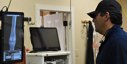
- Diagnostic imaging: Once the anatomic region of
interest is located, diagnostic imaging is used to
arrive at a diagnosis. A proper and accurate diagnosis
is absolutely essential to maximize the success of
treatment. Common diagnostic imaging modalities include
flouroscopy, digital radiology, and digital
ultrasonography. These technologies are non-invasive and
performed routinely prior to any advanced diagnostics.
When a diagnosis cannot be determined from these
modalities, advanced imaging modalities such as magnetic
resonance imaging (MRI) and nuclear scintigraphy (bone
scan) may need to be performed.
- Treatment: once an accurate diagnosis is made, a treatment plan must be formulated that is customized to the individual horse, keeping in mind the expectations of the owner and trainer, as well as the future competition schedule of the horse. Treatment options may include routine joint injection, corrective shoeing, shock wave therapy, IRAP therapy, surgery, or some sort of regenerative therapy (PRP or stem cell).
Common Conditions Responsible For Decreased Performance:
Suspensory Desmitis: Common in all disciplines in both the forelimb and hindlimb. Often appears in combination with other disorders, such as foot soreness in the forelimb and hock soreness in the hindlimb.
Stifle Disorders: Common in horses that utilize their hind ends, such as the western performance horse. Disorders can range from osteoarthritis to damage to soft tissue structures.Hock Disorders: Also common in the western performance horse as well as many other disciplines. Various stages of osteoarthritis is the most common diagnosis, but this may be in conjunction with other problems such as stifle and suspensory issues.
Foot Disorders: Common in all disciplines and age groups. Both front and rear limbs are affected. Generally a combination of joint, bone, and soft tissue disorders.
Cervical Spine (Neck) Disorders: Common in disciplines that work with collection. Osteoarthritis of the cervical facet joints is the most common diagnosis.
Thoracic and Lumbar Spine Disorders: Common in disciplines that work with collection. Can be a primary back problem such as osteoarthritis or muscle soreness secondary to a problem in one or multiple limbs.
Premier Equine Veterinary Services
130 Hughes Road / P. O. Box 1066
Whitesboro, Texas 76273
Phone/Fax: 855-HORSVET (855-467-7838)
[email protected]
Request Appointment